
Figure 4.
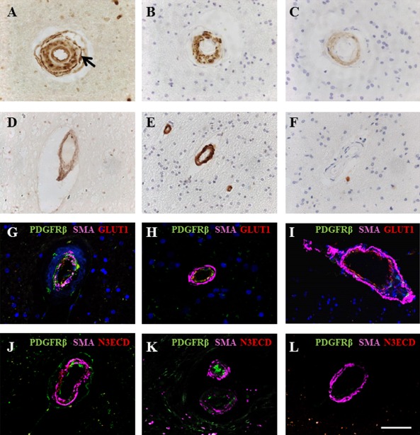
PDGFR‐β immunoreactivity associated with arterioles and cerebral microvessels in CADASIL. (A–C) Immunohistochemical staining for PDGFR‐β (A), SMA (B) and N3ECD (C) in a partially hyalinized arteriole in the white matter of a 44‐year‐old female CADASIL subject with R153C mutation. (A) PDGFR‐β reactivity was observed in the endothelial cells of arterioles and expressed abluminally to VSMC (arrow) and in the adventitia. (B) SMA and N3ECD reactivities were predominantly within the tunica media of the same vessel (C). (D–F) PDGFR‐β immunostaining in 49‐year‐old cognitively normal control female, indicating diffuse PDGFR‐β staining within arteriolar walls (D). SMA staining revealed intact vascular smooth muscle cells (E) without presence of N3ECD aggregates (F). (G–I) Immunofluorescent labelling of PDGFR‐β (green), SMA (magenta) and GLUT‐1 (red) in arterioles of the frontal white matter, with DAPI counterstain. (G) Vascular smooth muscle cell (demonstrated by SMA, magenta) degeneration in arterioles in a 68‐year‐old female CADASIL case with R133C mutation, with increased PDGFR‐β (green) expression within the vessel wall. (H) Some PDGFR‐β (green) reactivity in the endothelial cell layer of arteriole from an 81‐year‐old female patient with sporadic SVD with mild VSMC degeneration compared with CADASIL (cf. D). (I) PDGFR‐β (green) expression was absent in an arteriole in a 94‐year‐old nondemented female control, although GLUT‐1 (red) and SMA (magenta) appeared normal. (J–L) Immunofluorescent labelling of PDGFR‐β (green), SMA (magenta) and N3ECD (red) in arterioles of white matter, confirming that N3ECD only accumulated in the tunica media in CADASIL (J), although PDGFR‐β (green) immunoreactivity was apparent in both arterioles from CADASIL (J) and SVD cases (K). (L) GLUT‐1 immunostaining in endothelial cells of arteriole of a control subject, whereas PDGFR‐β reactivity was meagre, if any. Scale bar = 50 μm in A–F, 40 μm in G, 27.5 μm in H, 47.5 μm in I, 52 μm in J, 57 μm in K and 49.5 μm in L. CADASIL, cerebral autosomal dominant arteriopathy with subcortical infarcts and leukoencephalopathy; PDGFR‐β, platelet‐derived growth factor receptor‐β; SMA, smooth muscle alpha actin; SVD, small vessel disease; VSMC, vascular smooth muscle cell.