
Figure 6.
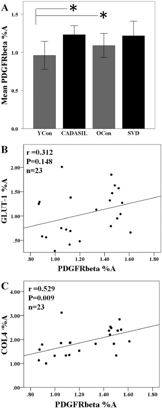
Quantification of PDGFR‐β immunoreactivity in the frontal white matter in CADASIL. (A) Quantitative analysis of PDGFR‐β percent area (%A) revealed a trend for differences between the groups (anova, P = 0.084, n = 6–7 samples per group). However, post hoc tests revealed significant differences between CADASIL subjects and young controls, and the old control group compared with young controls (Tukey's test, *P < 0.05). (B) Lack of relationship between PDGFR‐β %A and GLUT‐1 immunoreactivity (r = 0.312, P = 0.148). (C) PDGFR‐β %A was significantly correlated with the basement membrane marker COL4 (r = 0.529, P = 0.009). CADASIL, cerebral autosomal dominant arteriopathy with subcortical infarcts and leukoencephalopathy; PDGFR‐β, platelet‐derived growth factor receptor‐β.