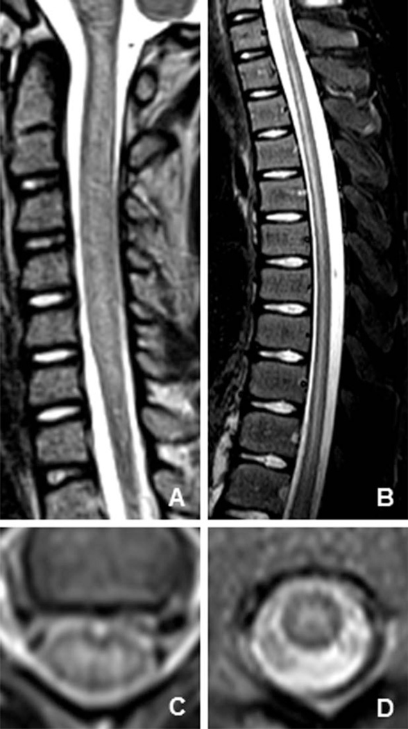
FIGURE 2.
Representative magnetic resonance imaging (MRI) images of the spinal cord in acute flaccid myelitis cases in the United States 2012–2015. MRI images from acute flaccid myelitis patients in the Children’s Hospital Colorado cohort. (A and B) Saggital T2-weighted MRI sequences of the spinal cord demonstrate longitudinally extensive, hyperintense lesions. (C and D) Axial T2-weighted MRI sequences of the spinal cord demonstrate predominant involvement of the spinal cord gray matter.

