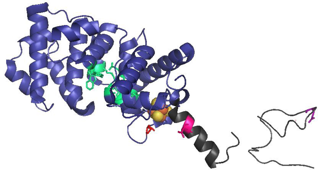
Figure 2.
Locations of MUTYH amino acid variations. The X-ray crystal structure the N-terminal domain of MUTYH (residues 65–350) (PDB code 3N5N) illustrates the amino acid positions of P281L, R295C and Q324H MUTYH variants. The N-terminal catalytic domain is shown in blue, the adenine-specific active site pocket in green, the [4Fe-4S]2+ cluster in orange/gold spheres and the IDC/linker region in dark grey. The locations of the WT amino acids Pro 281 (red), Arg295 (pink) and Gln324 (purple) are highlighted in stick form on the ribbon backbone.

