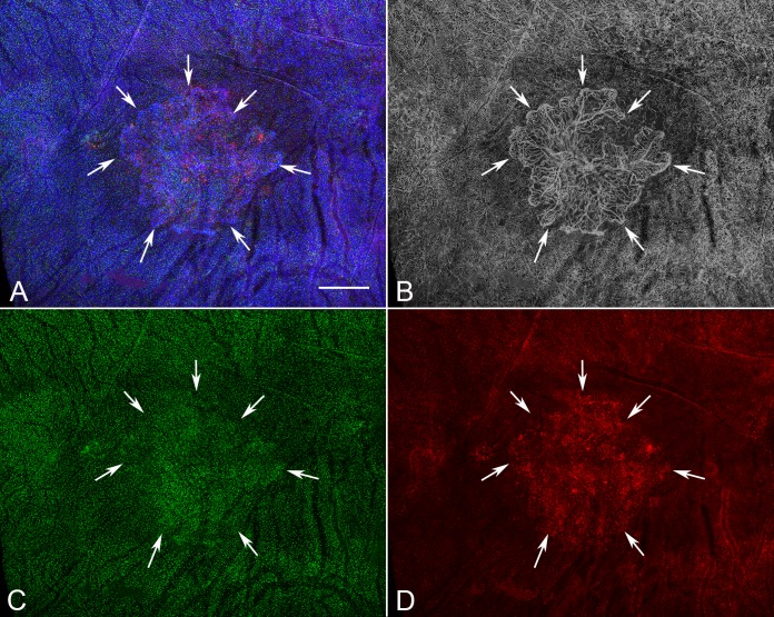
Figure 9.
Low-magnification image of the posterior pole region of choroid from a subject (subject 14, OS) with neovascular AMD showing a CNV membrane in the submacular region (arrows). There was an increase in both IBA1+ (C) and HLA-DR+ macrophages (D) associated with the CNV in neovascular AMD eyes. (A) Merged channels; (B) desaturated UEA channel; (C) IBA1 channel; (D) HLA-DR channel. Scale bar: 1 mm.

