Figure 5.
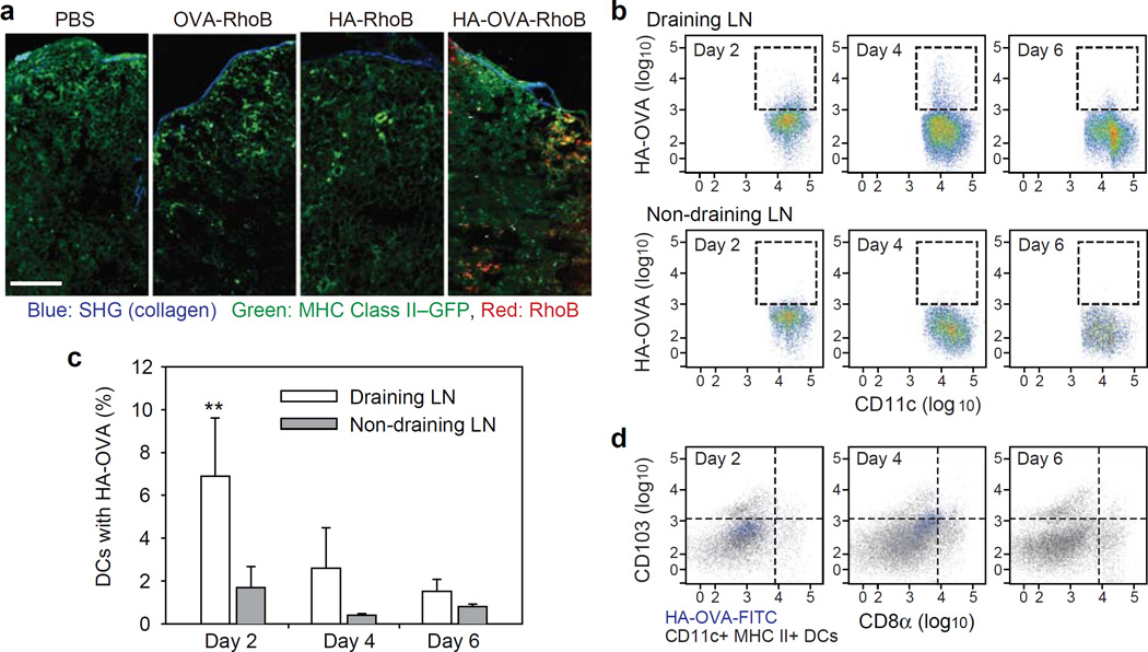
(a) Two-photon microscopic image of histological sections of skin-draining LNs 2 days after treatment with PBS, OVA-RhoB, HA-RhoB, and HA-OVA-RhoB conjugates, respectively, on the back skin of mice. Scale bar, 100 μm. (b) Cytometry plots of HA-OVA-FITC conjugate containing CD11c+ DCs in skin draining LNs (Top) and non-draining LNs (Bottom), at 2, 4 and 6 days post topical application on abdominal flank. (c) The ratio of CD11+ DCs containing HA-OVA-FITC conjugate determined from the cytometry results (mean ± SD, n = 5). **, P < 0.01, Draining LN vs. Non-draining LN. (d) Cytometry analysis for the expression of CD8α and CD103 of the cells associated with HA-OVA-FITC conjugate or CD11c+ MHC II+, at 2, 4, and 6 days in draining inguinal LNs.

