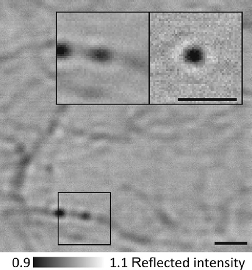
Fig. 2.
iSCAT assay for gold-labeled myosin-5a tracking. Flat-field-corrected image showing actin filaments and some 20 nm gold-labeled myosin-5a molecules bound to actin. Inset: zoom of the indicated region showing 20 nm gold-labeled myosin-5a bound to actin (left) and the same region after background subtraction, which removes all static features including the actin filament and any immobile particles (right). Scale bars: 1 µm.

