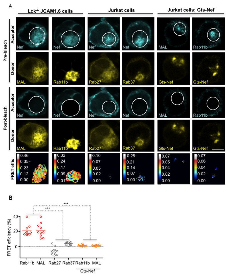
Figure 3. Nef selectively associates with the regulators of Lck vesicular traffic Rab11b and MAL.
(A) FRET imaging of Lck-deficient JCAM1.6 or Jurkat vesicular compartments by acceptor photobleaching. Top, imaging prior to photobleaching of the mCherry-tagged FRET-acceptors (Pre-bleach; Acceptor) and of the GFP-tagged FRET-donors (Pre-bleach; Donor). Middle, imaging post photobleaching of the mCherry-tagged FRET-acceptors (Post-bleach; Acceptor) and of the GFP FRET-donors (Post-bleach; Donor). Bottom panels display calculated [(Donorfinal-Donorinitial)/Donorfinal] FRET efficiency maps. Bleached areas are indicated by white circles.
(B) Population analysis of FRET efficiencies for n=10 per experimental condition.
Each symbol represents a cell. Scale bar, 5 μm. Data are representative (A, B, D) or represent (C, E) 2 independent experiments ***P ≤ 0.0001; n.s. not significative (Mann-Whitney test).

