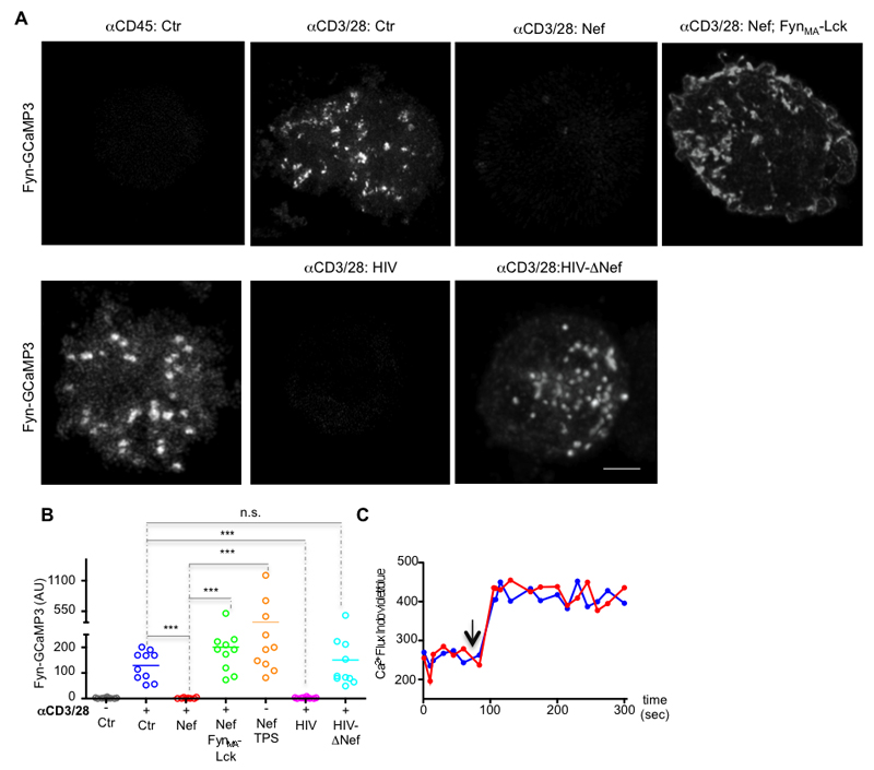
Figure 5. Nef impairs the formation of Ca2+ territories at the synaptic membrane.
(A) The membrane tethered and genetically encoded Ca2+ indicator (Fyn-GCaMP3) was used to image the formation of Ca2+ territories at the synaptic membrane of control, Nef expressing (Nef), Nef and FynMA-Lck co-expressing (Nef; FynMA-Lck), HIV or HIV-ΔNef infected Jurkat cells while interacting with glass coated with antibodies against CD3 and CD28 (αCD3/28) or with αCD45 (αCD45; Ctr) for 3 min before imaging.
(B) Population analysis of the formation Ca2+ membrane domains at the immunological synapse in Jurkat cells processed as in (A) (arbitrary units).
(C) Cytosolic Ca2+ flux measurements (showing the ratio of indo-blue to indo-violet) in control (blue) and Nef-expressing (red) Jurkat cells stimulated with ionomycin (arrow).
Each symbol represents a cell. Scale bar 5 μm. Data are representative (A, C) or represent (B) 3 independent experiments ***P ≤ 0.0001; n.s. not significative (Mann-Whitney test).

