Fig. 1.
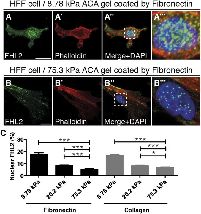
FHL2 localization depends on substrate rigidity. Confocal immunofluorescence images of FHL2, actin, and nuclear staining in HFF cells cultured on ACA gels of different rigidities for 17 h. FHL2 localization was labeled by the FHL2 antibody (green), actin filaments were stained with Alexa Fluor 594 phalloidin (red), and the nucleus was counterstained with DAPI (blue). (Scale bars: 20 μm.) (A–A′′) HFF cells on an 8.78-kPa ACA gel coated with fibronectin. (A′′′) Zoomed-in view of the dotted box in A′′, showing nuclear localization of FHL2 and actin. (B–B′′) HFF cell on a 75.3-kPa ACA gel coated with fibronectin. (B′′′) Zoomed-in view of the dotted box in B′′, showing nuclear localization of FHL2 and actin. (C) Graph showing the intensity ratio of FHL2 immunofluorescence staining between the nucleus and whole cell area (nucleus/whole cell area), fibronectin- or collagen-coated gels of varying rigidity. All images are shown as projected images from adhesion sections to nuclear sections. n > 30. Error bars represent SEM. *P < 0.05; ***P < 0.0001.

