Fig. 4.
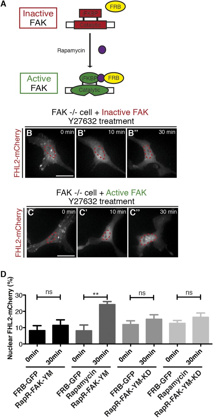
Kinase activity in FAK is responsible for FHL2 transport to the nucleus. (A) Schematic image illustrating the allosteric activation of RapR-FAK by rapamycin treatment (47). (B–B′′) FHL2-mCherry dynamics in FAK−/− cells expressing RapR-FAK-YM and FRB-GFP expression (inactive FAK) after Y-27632 treatment. (C–C′′) FHL2-mCherry dynamics in FAK−/− cells with RapR-FAK-YM and FRB-GFP expression and rapamycin treatment (active FAK) after Y-27632 treatment. (D) Graph showing the intensity ratio of FHL2-mCherry in FAK−/− cells between nuclei and whole cells using the RapR-FAK system before and after Y-27632 treatment. The magenta circle indicates NLS-BFP (nuclear marker). (Scale bars: 20 μm.) All images are projected images from adhesion sections to nuclear sections. n > 15. Error bars represent SEM. **P < 0.001.

