Fig. 5.
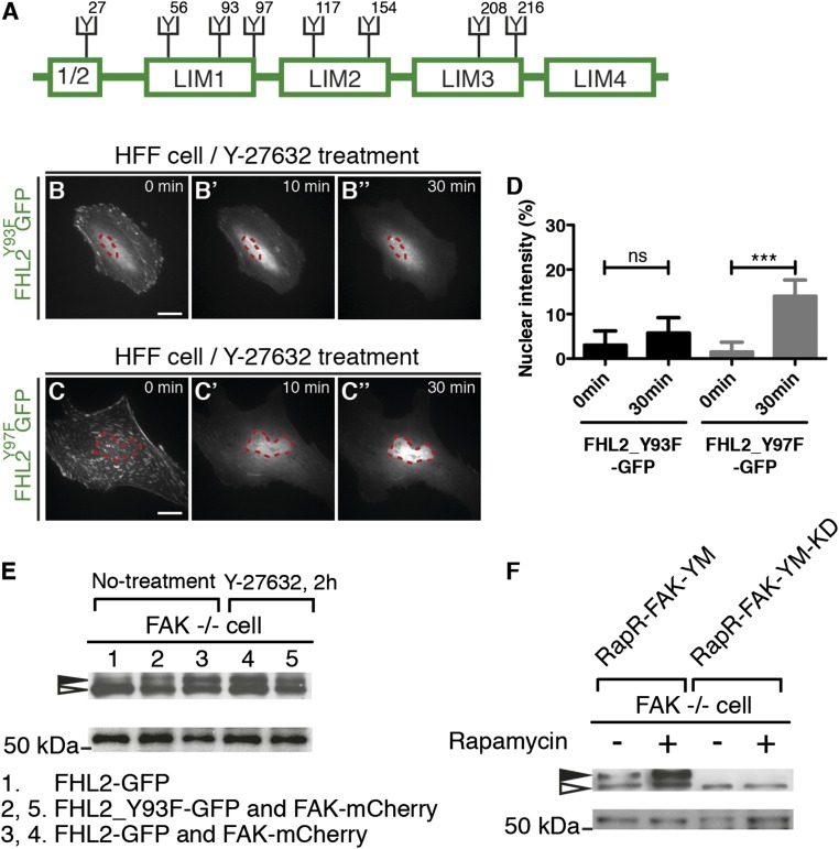
The tyrosine-93 residue in FHL2 is responsible for FHL2 nuclear transport. (A) Schematic image showing the location of each tyrosine residue in FHL2. (B–B′′) The dynamics of FHL2-GFP with tyrosine-93 mutation (Y93F) in HFF cells after Y-27632 treatment. (C–C′′) The dynamics of FHL2-GFP with tyrosine-97 mutation (Y97F) in HFF cells after Y-27632 treatment. (D) Graph showing the intensity ratio of FHL2_Y93F-GFP or FHL2_Y97F-GFP between nuclei and whole cells after Y-27632 treatment. (Scale bars: 20 μm.) (E, Top) SDS/PAGE with polyacrylamide containing Mn2+ and Phos-tag. Black arrow, phosphorylated FHL2; white arrow, unphosphorylated FHL2. (E, Bottom) SDS/PAGE with polyacrylamide. Western blot analysis for FHL2-GFP or FHL2_Y93F-GFP from each sample isolated from FAK−/− cells and FAK−/− cells rescued by FAK-mCherry. Lane 1, FHL2-GFP from FAK−/− cells; lane 2, FHL2_Y93F_GFP from FAK−/− cells rescued by FAK-mCherry; lane 3, FHL2-GFP from FAK−/− cells rescued by FAK-mCherry; lane 4, FHL2-GFP from FAK−/− cells rescued by FAK-mCherry with Y-27632, lane 5; FHL2_Y93F-GFP from FAK −/− cell rescued by FAK-mCherry with Y-27632. (F, Top) SDS/PAGE with polyacrylamide containing Mn2+ and Phos-tag. Black arrow, phosphorylated FHL2; white arrow, unphosphorylated FHL2. (F, Bottom) SDS/PAGE with polyacrylamide. Western blot analysis for FHL2-GFP from each samples isolated from FAK−/− cells with RapR-FAK-YM or RapR-FAK-YM-KD and myc-FRB expression, with and without rapamycin treatment. (F, Left) FHL2-GFP from FAK−/− cells with RapR-FAK-YM and myc-FRB expression, without and with rapamycin treatment. (F, Right) FHL2-GFP from FAK−/− cells with RapR-FAK-YM-KD and myc-FRB expression, without and with rapamycin treatment. (Scale bars: 20 μm.) All images are projected images from adhesion sections to nuclear sections. n > 10. Error bars represent SEM. ***P < 0.0001.

