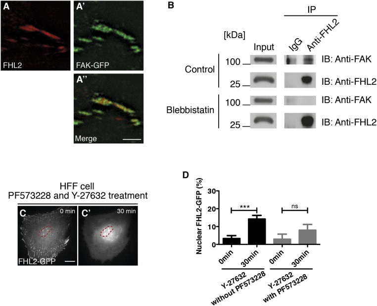
Fig. S7.
FHL2 interacts with FAK in HFF cells. (A–A′′) Images obtained with a Nikon N-SIM superresolution microscope. (A) Superresolution image of FHL2 localization at the adhesion in HFF cells. (A′) Superresolution image of FAK-GFP localization at the adhesion in HFF cells. (A′′) Merged image of A and A′. (B) Western blot analysis for FAK with the cell lysate from HFF cells after IP with FHL2 antibody with or without blebbistatin. (C and C′) FHL2-GFP dynamics after PF573228 (10 μM, 7 h) and Y-27632 treatment. (D) Graph showing the relative intensity of FHL2-GFP at the nucleus in HFF cells after PF573228 and Y-27632 treatment. n > 20. Error bars represent SEM. ***P < 0.0001. The magenta circle indicates NLS-BFP (nuclear marker).

