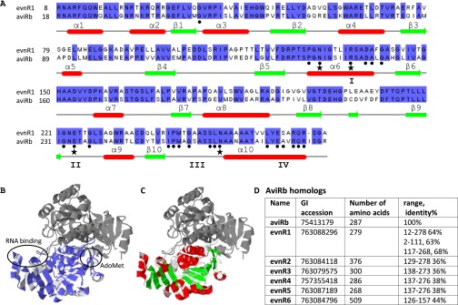
Fig. S4.
Sequence alignment of evnR1 and aviRb superposed on aviRb structure. (A) The conserved nucleotides are highlighted in purple. (B) Secondary structure elements of aviRb are given for reference in red and green. (C) AviRb crystal structure (1 × 7O, 1 × 7P). The RNA-binding region of the N-terminal domain was derived from the structurally homologous ribosomal proteins L30 and L7Ae; it is marked in purple. The alignment is based on aviRb homologs, given in D.

