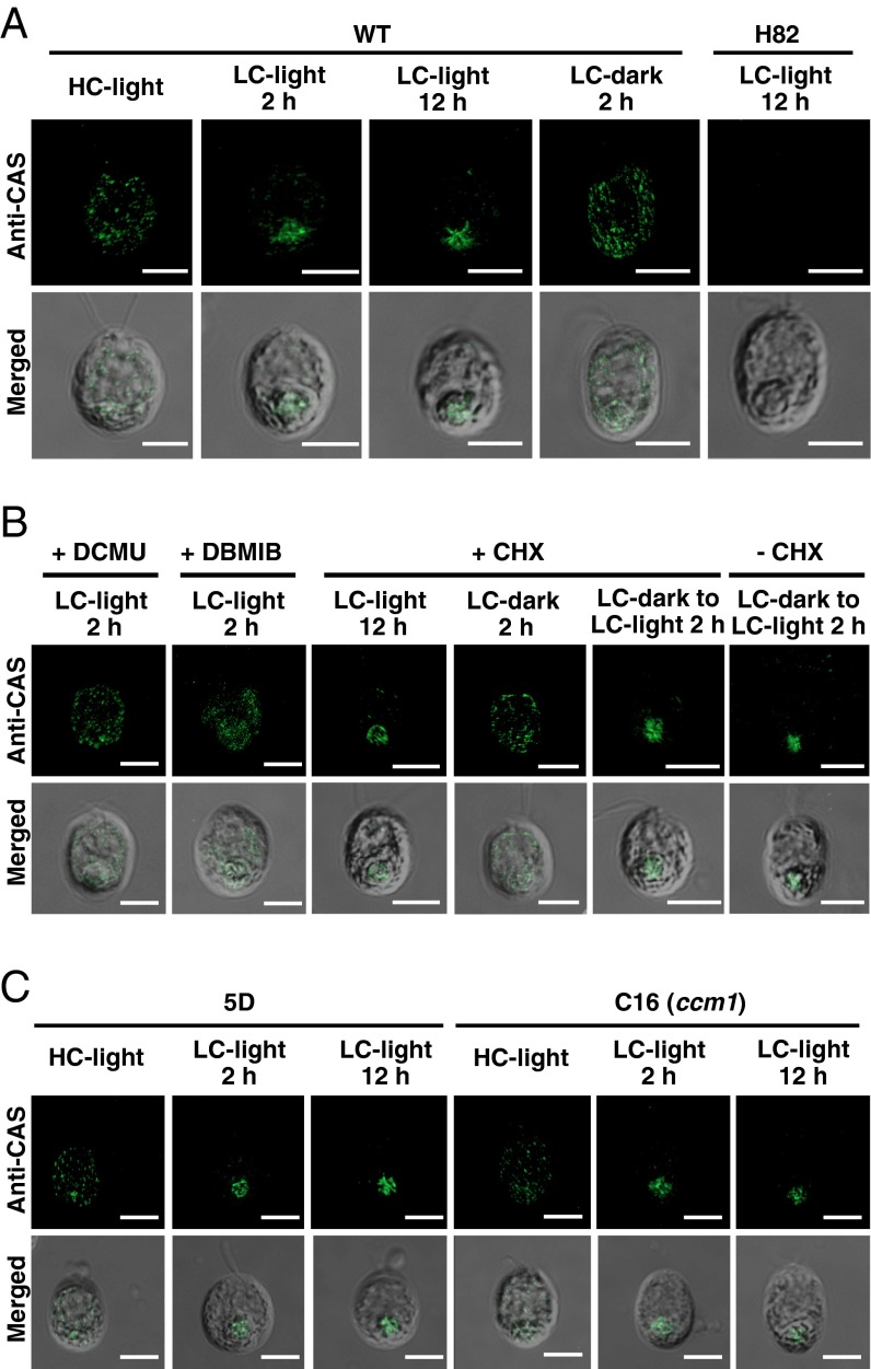
Fig. 3.
Subcellular localization of CAS. (A) Localization of CAS in WT and H82 cells was assessed using an indirect immunofluorescence assay with an anti-CAS antibody. Cells grown in high-CO2 (HC) conditions were shifted to low-CO2 (LC) conditions for 2 h or 12 h in light, and then the LC-acclimated cells (12 h) were transferred from light to dark for 2 h in the LC condition. (B) The effect of dichlorophenyl-dimethylurea (DCMU) (10 µM), 2,5-dibromo-3-methyl-6-isopropylbenzoquinone (DBMIB) (10 µM), or cycloheximide (CHX) (10 µg⋅mL–1) on the localization of CAS in WT cells. The cells cultured in each condition used in A were also used for these drug treatments. The HC-grown cells were incubated under LC-light conditions in the presence of DCMU or DBMIB for 2 h. For CHX treatments, HC-grown cells were transferred to LC-light conditions in the presence of CHX for 12 h. The LC-light–acclimated cells were subjected to LC-dark conditions in the presence of CHX for 2 h. The LC-dark–acclimated cells were transferred to LC-light conditions with or without CHX for 2 h. (C) Localization of CAS in a CCM1 insertion mutant, C16, and its parental 5D strain using an indirect immunofluorescence assay. Cells grown in HC conditions were shifted to LC-light conditions for 2 h or 12 h. For all panels, each image is placed with the flagella facing upward on the panel. Merged images show the fluorescence image superimposed with its differential interference contrast image. (Scale bars: 5 µm.)

