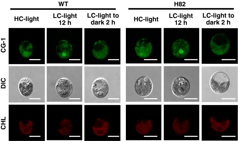
Fig. 4.
Calcium Green-1, AM fluorescence in WT and H82 cells. Cells were grown in high-CO2 (HC) or low-CO2 (LC) conditions for 12 h in light, or cells in LC-light were transferred to LC-dark conditions for 2 h, and then incubated with Calcium Green-1, AM (CG-1) at room temperature for 30 min. Fluorescence images derived from CG-1 treatments and chlorophyll (CHL) are shown. Each image is placed with the flagella facing upward on the panel. DIC, differential interference contrast image. (Scale bars: 5 µm.)

