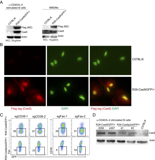
Fig. S3.
Cas9 levels and localization in primary cells of R26-Cas9iGFP/+ mice and Cas9-dose-dependent knockout efficiency. (A) Western blots of Cas9 proteins (detected by anti-Cas9 and anti-Flag antibodies) in anti-CD40/IL-4–activated B cells and BMDMs isolated from R26-Cas9iGFP/+ and wild-type animals. β-Actin was used as a loading control. (B) Immunostaining of Cas9 in BMDMs derived from R26-Cas9iGFP/+ and wild-type animals. Cas9 was stained using anti-Flag (M2) antibodies (red). DAPI was used for counterstaining of nuclei (green). (C) Knockout efficiencies in B cells from R26-Cas9iGFP/+ (Top) and R26-Cas9p2aGFP/+ (Bottom) mice that were activated and transduced with retroviruses expressing sgCD38-1, sgCD38-2, sgFas-1, and sgFas-2 side by side. The gates in the FACS plots show the percentage of cells that lost the surface marker 4 d after transduction. (D) Western blots of Cas9 proteins in anti-CD40/IL-4–activated B cells isolated from R26-Cas9iGFP/+, R26-Cas9p2aGFP/+, and wild-type animals. The data are representative of two independent experiments.

