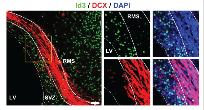
Figure 2.

Id3 is not expressed in DCX+ type A neuroblasts in the RMS. Representative image of uninjured WT mice immunolabeled for Id3 (green) and DCX (red, marker for type A neuroblasts). Nuclei are stained with DAPI (blue). LV, lateral ventricle; RMS, rostral migratory stream; SVZ, subventricular zone. Scale bar: 50 µm.
