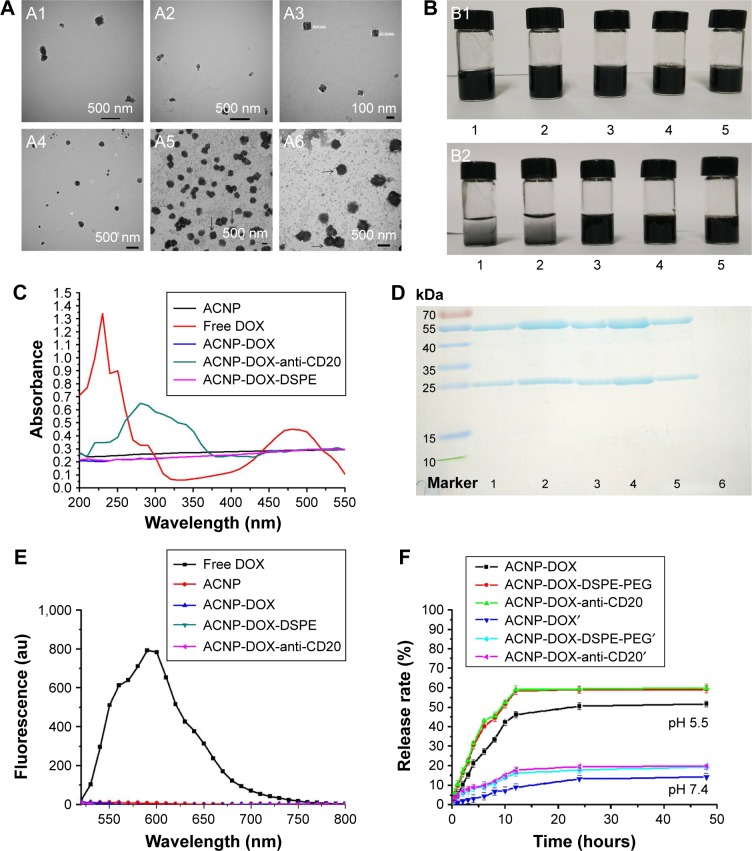
Figure 2.
ACNP-DOX-DSPE-PEG2000-anti-CD20 NDDS characterization.
Notes: (A) The images of TEM show that modification of ACNP changed the morphology of the formulation. ACNP (A1) and ACNP-DOX (A2) particles were irregular in shape. ACNP-DOX-DSPE-PEG2000 (A3) particles had a cubic morphology with good dispersion stability. ACNP-DOX-DSPE-PEG2000-anti-CD20 (A4) particles were spherical in shape. There were no projections on the stained cubic particles of ACNP-DOX-DSPE-PEG2000, and they are indicated by black arrows (A5). However, cilia-like projections could be seen on the surface of the stained spherical particles of ACNP-DOX-DSPE-PEG2000-anti-CD20, and they are indicated by black arrows (A6). The samples of A1, A2, A3, and A4 were not negative-stained during preparation process, and A5, A6 were stained with 1% uranyl acetate. (B) The dispersion and stability of blank ACNP (1), ACNP-DOX (2), ACNP-DOX-DSPE-PEG2000 (3), ACNP-DSPE-PEG2000 (4), and ACNP-DOX-DSPE-PEG2000-anti-CD20 (5) in pH 7.4 PBS. The solutions were put at 4°C for over 1 month. ACNPs had aggregated and settled to the bottom of the ampule in the ACNP and ACNP-DOX groups, but no precipitates were seen at the bottom of containers in ACNP-DSPE-PEG2000, ACNP-DOX-DSPE-PEG2000 and ACNP-DOX-DSPE-PEG2000-anti-CD20 groups (B2). The original concentration of them was 0.5 mg/mL. (C) UV–Vis absorption spectra of DOX, ACNP, ACNP-DOX and ACNP-DOX-DSPE-PEG2000-anti-CD20. The wavelength of maximum absorption peak was 480 nm for free DOX, and 254 nm for ACNP-DOX-DSPE-PEG2000-anti-CD20. There were no maximum absorption peaks for the other three samples. (D) SDS-PAGE analysis showed that the first, second, third, and fourth lanes included the big and small subunits of free anti-CD20 antibodies; the fifth lane included the subunits of anti-CD20 in ACNP-DOX- DSPE-PEG2000-anti-CD20; the sixth lane had no subunits of anti-CD20 in ACNP-DOX-DSPE-PEG2000 (ACNP unconjugated from anti-CD20). (E) Fluorescence spectra of DOX, ACNP, ACNP-DOX and ACNP-DOX-DSPE-PEG2000-anti-CD20. Only free DOX was found to have a strong fluorescence at 590 nm. (F) The in vitro release profile of DOX from ACNP-DOX, ACNP-DOX-DSPE, and ACNP-DOX-DSPE-antiCD20 with similar cumulative release behavior in pH 7.4 and 5.5 PBS. The release rates of DOX were no more than 20% in pH 7.4 PBS at 37°C during 48 hours, but at pH 5.5 they were 60% within 12 hours.
Abbreviations: au, astronomical unit; ACNP, active carbon nanoparticles; DOX, doxorubicin; DSPE, 1,2-distearoyl-sn-glycero-3-phosphoethanolamine; PEG, polyethylene glycol; UV–Vis, ultraviolet–visible.

