Figure 4.
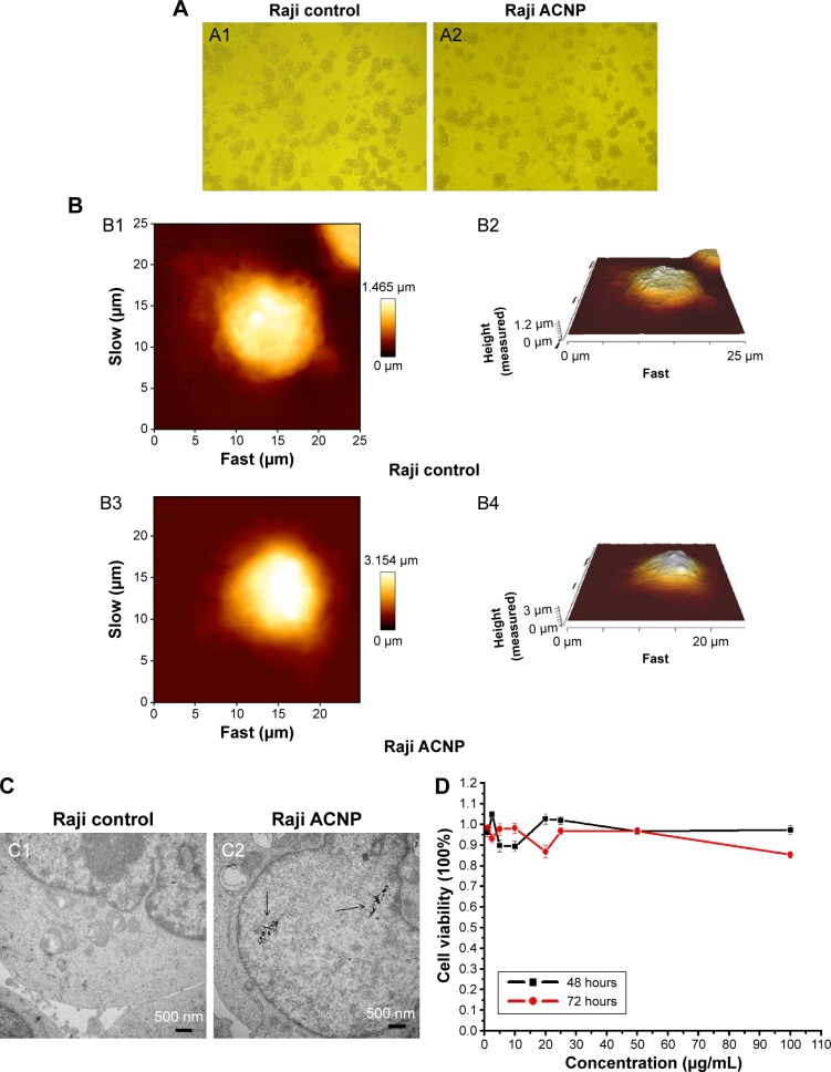
The cytotoxicity of ACNP on Raji cells (A) light microscopy (A1 and A2, ×100) (B) AFM (the image sizes of Bl and B3 are 25×25 μm, B2 and B4 are their corresponding three-dimensional structures) (C) TEM (D) MTT.
Notes: (A) The light microscopic morphology of Raji cells cultured in 20 μg/mL ACNP for 72 hours, with comparison of the untreated Raji cells (A1) and the treated Raji cells (A2) (A1 and A2, ×100). (B) AFM showed that the surface of the Raji cells in the control group (B1 and B2) showed no abnormalities when compared with the ACNP-treated group (B3 and B4) (the image sizes of B1 and B3 are 25 μm×25 μm, B2 and B4 are their corresponding 3D structures). (C) TEM demonstrated no differences in the ultrastructure of the nucleus and organelles between the untreated (C1) and ACNP-treated cells (C2), although ACNPs were demonstrated to enter the cell (black arrows in C2). (D) The MTT assays showed that treatment with ACNP did not inhibit the growth of Raji cells even at the highest concentration (100 μg/mL), indicating that ACNP may be a potential drug delivery carrier that is without immediate cytotoxicity.
Abbreviations: AFM, atomic force microscopy; ACNP, active carbon nanoparticles; μm, micrometer; MTT, 3-(4,5-Dimethylthiazol-2-yl)-2,5-diphenyltetrazolium bromide; TEM, transmission electron microscope.

