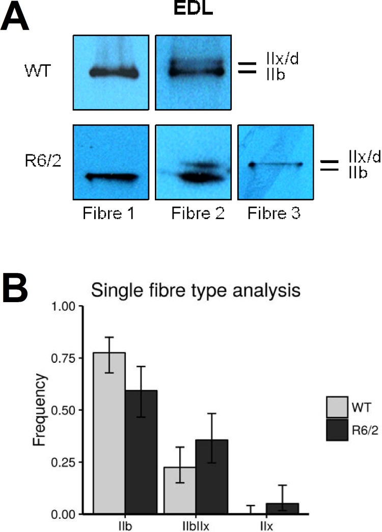
Fig 3. MyHC determination in single muscle fibers.
Muscle fiber composition for EDL was assessed on a single fiber basis. For this MyHC were extracted from 6 to 15 randomly selected, intact fibers per muscle sample. (A) Examples showing Roti®-Blue-stained MyHC bands from selected single fiber SDS PAGE gels. Fibers were classified as Type IIB and IIX when a single band could be detected and as mixed IIB/X when both bands were detectable, irrespective of staining intensity. (B) Relative contribution (fractional number) of pooled fibers (59 fibers of 5 R6/2 mice and 89 fibers of 7 WT mice) exhibiting expression of MyHC IIb, IIx or both. Distributions of fibers amongst the types IIB, IIX and mixed type IIB/X differed significantly between WT and R6/2 mice (p = 0.02) indicating a different muscle fiber composition of R6/2 EDL muscle with more mixed-type fibers. Bars show relative frequencies; error bars show confidence intervals for binomial proportions (i.e. fibers of respective type vs. all other fibers) (95% CI).

