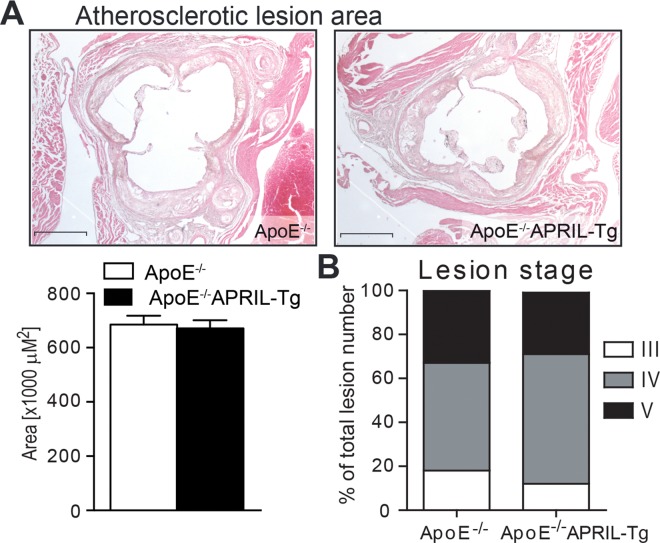
Fig 2. Lesion size and stage of ApoE-/- and ApoE-/-APRIL-Tg mice.
After 12 weeks of WTD lesion size (A) was quantified and lesion stage (B) was determined in the aortic roots of ApoE-/- (n = 13) and ApoE-/-APRIL-Tg mice (n = 10). Representative photomicrographs are shown with original magnification x25. Data are represented as mean±SEM; Scale bars represent 1mm.

