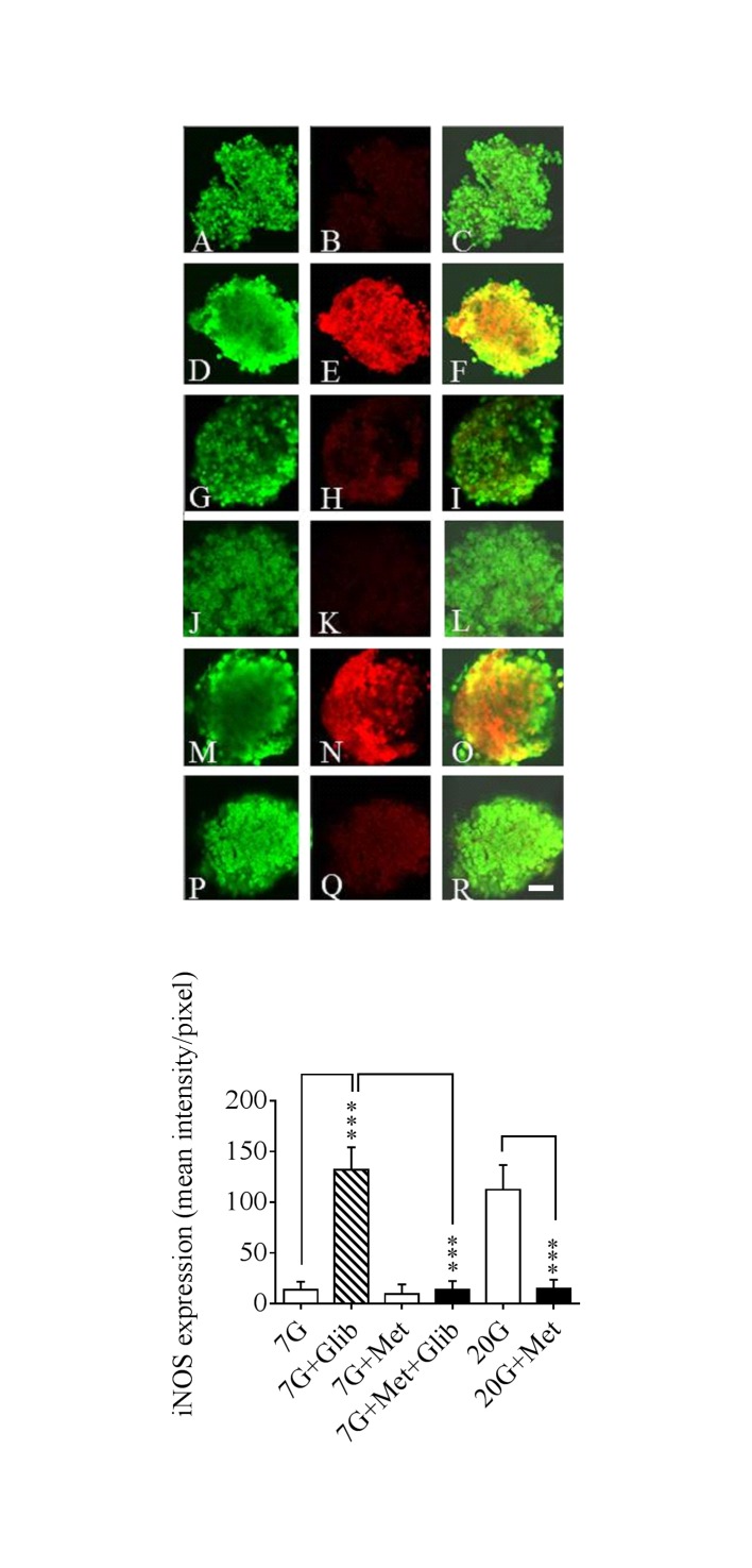
Fig 3. Confocal microscopy and fluorescence intensity after 24 h culturing.
Confocal microscopy and fluorescence intensity measurements of murine islets cultured for 24h at 7 mmol/l glucose (7G) ± 5 μmol/l glibenclamide (Glib) or 20 μmol/l metformin (Met) or both as well as at 20 mmol/l glucose (20G) ± 20 μmol/l metformin (Met). The islets were double-immunolabelled for insulin (A, D, G, J, M, P) and iNOS protein (B, E, H, K, N, Q). Insulin staining appears as green and iNOS as red staining, respectively. Co-localization of insulin/iNOS is seen as orange-yellowish fluorescence (C, F, I, L, O, R). Bars indicate 20 μm Lower part of the figure shows fluorescence intensity measurements quantified pixel by pixel using Zen 2009 software. Means ± SEM are shown for 19–34 observations in each group. ***p<0.001

