
Figure 1.
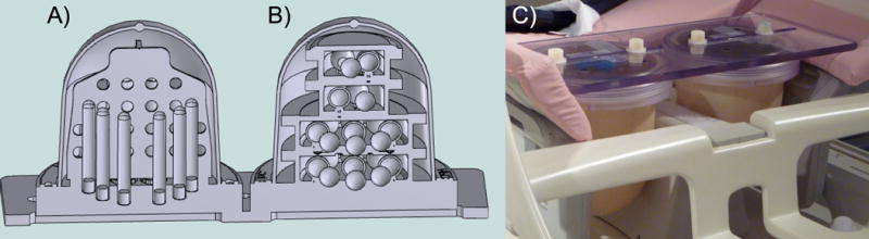
Breast phantom CAD model showing A) the diffusion phantom unit with vertically oriented samples tubes and geometric distortion plate and B) the T1 phantom unit with sample spheres arranged on four isolated levels, and C) prototype prepared for imaging in a breast MR coil.