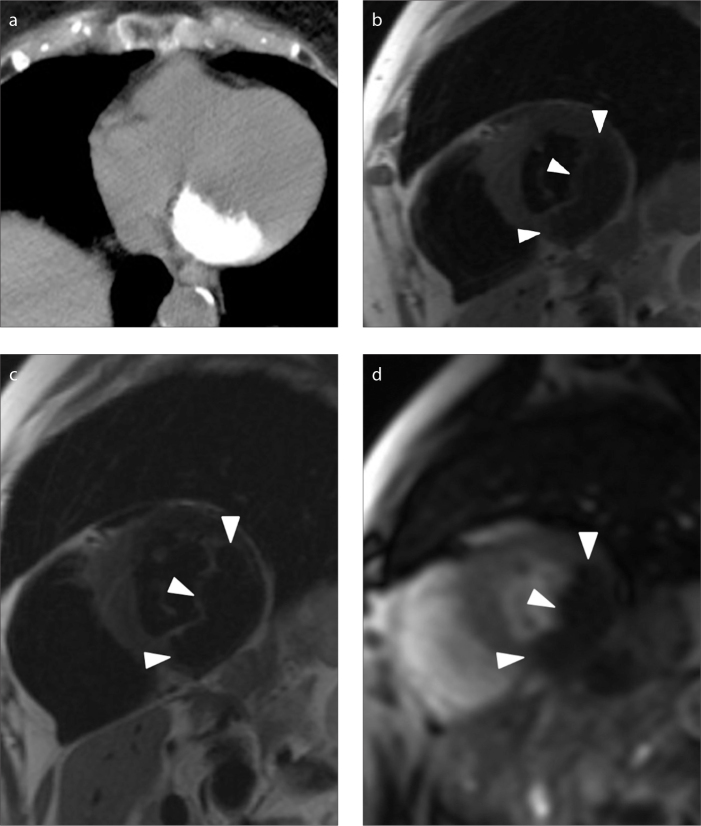
Figure 4.
a–d. An asymptomatic 87-year-old woman with a 37×14 mm polypoid mass with a broad-based appearance (case no: 11). CT image (a) shows a diffuse calcified lesion. Short axis T1-weighted (b) and T2-weighted (c) black blood images show a hypointense mass (arrowheads) compared with the signal intensity of the myocardium. Short axis T1-weighted image after gadolinium administration (d) shows no enhancement of the mass.

