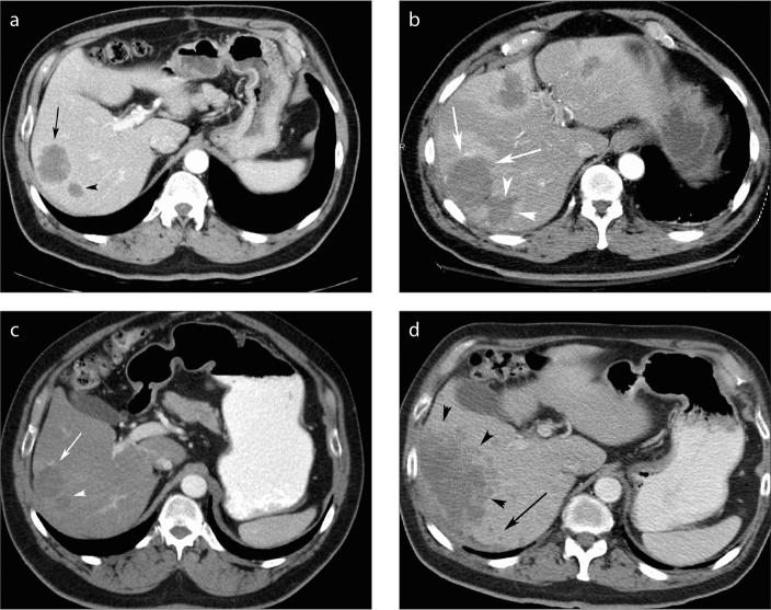
Figure 2.
a–d. Example of circumferential local progression of disease in a 67-year-old man on chemotherapy for CRCLM. Axial contrast-enhanced CT six weeks pre-RF ablation (a) demonstrates two lesions next to each other in the right lobe of the liver (black arrow and black arrowhead). CT performed one month post-RF ablation (b) demonstrates primary technical success with ablation margins measuring >5mm compared with the original tumors (white arrows and arrowheads). Two extra areas of ablation were performed in this patient since two new hypodense lesions were identified at the time of the RF ablation in segments 4 and 2/3 of the liver (not shown). Three months post-RF ablation (c), the ablated areas had reduced in size and were similar in size to the original tumors (white arrow and arrowhead). The patient’s condition deteriorated and a large area of local progression of disease was noted around the entire margin of the previously treated lesion (d, black arrowheads). In addition, new subcentimeter hypodense satellite lesions (black arrow) can be seen. The aggressiveness of the patient’s underlying malignant disease may have contributed to the development of local progression of disease and satellite lesions.

