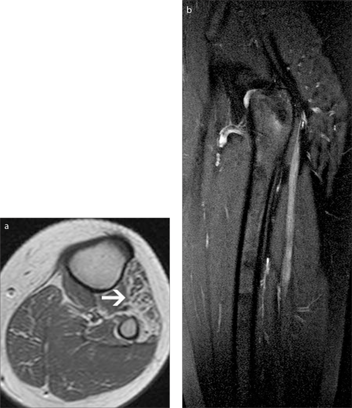
Figure 10.
a, b. An 11-year-old boy with a complaint of weakness of the ankle dorsiflexion and evertion, which yielded a peroneal nerve compromise. Axial T1-weighted image (a) of the knee shows fatty atrophy indicating chronic denervation of the lateral compartment muscles (arrow). As the common peroneal nerve was extending uneventfully, MRI of the SN was recommended. On sagittal fat-saturated T2-weighted image (b), the SN is thickened with preservation of the fascicles seen as tiny hypointense dots on axial fat saturated T2-weighted images (not shown). Findings are compatible with localized hypertrophy of the SN in this case.

