Figure 13.

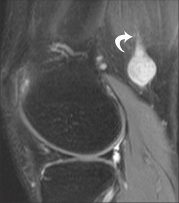
A 47-year-old male with schwannoma. Sagital fat saturated T2-weighted image shows a well-defined hyperintense mass in the posterior supracondylar region of the knee, with the SN entering the mass (curved arrow).

A 47-year-old male with schwannoma. Sagital fat saturated T2-weighted image shows a well-defined hyperintense mass in the posterior supracondylar region of the knee, with the SN entering the mass (curved arrow).