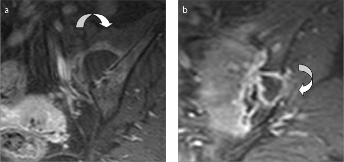
Figure 4.
a, b. A 51-year-old female with a history of epidural analgesia. Oblique-coronal fat-saturated T1-weighted contrast-enhanced (intravenous gadolinium-based agent) images show septic sacroiliitis on her left sacroiliac joint with multiple abscess formations (arrows) extending to presacral region (a) and posteriorly to gluteal muscles (b). The abscesses lying adjacent to the SN adds to her severe lower back pain. Bacterial

