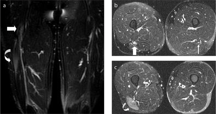
Figure 9.
a–c. A 26-year-old male with a history of sharp cut wound to his right thigh, who presented with pain and plantar flexion weakness. Coronal (a) and axial (b, c) fat-saturated T2-weighted images of both thighs show fusiform enlargement of the SN (thick arrows) with nerve continuity about the level of the previous trauma (a, b). The axial plane on panel (b) corresponds to the level where the fusiform enlargement of the nerve is shown on panel (a). Compare the thickened right-sided SN with the normal left side (thin arrow) (b). Denervation edema (curved arrow) in the long head of the biceps femoris muscle (a, c) is seen with diffuse hyperintensity in the muscle.

