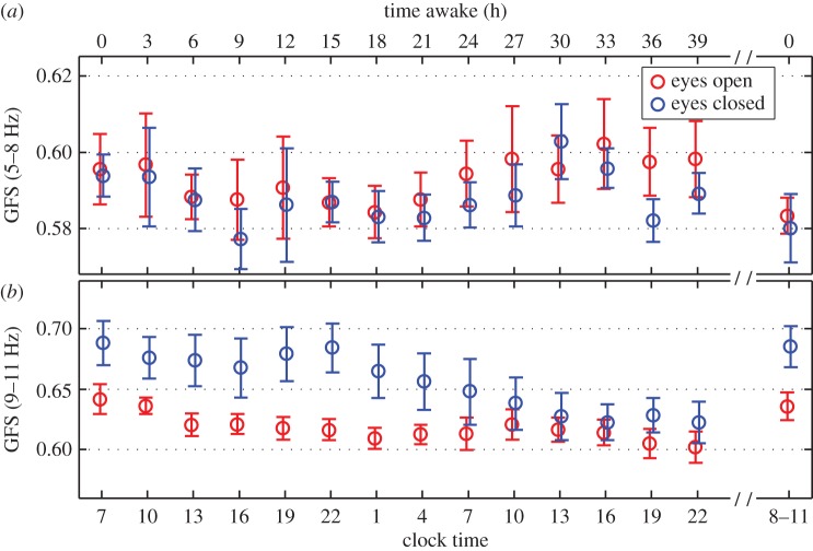
Figure 4.
Time course of GFS in the theta (5–8 Hz) and alpha (9–11 Hz) band of the wake EEG during sustained wakefulness of 40 h. Time awake (a) and clock time (b) are indicated. The last data points are after a night of recovery sleep following sleep deprivation. GFS decreased as a function of sleep deprivation in the eyes closed condition only. We note that one subject did not contribute to the 9 h after waking (clock time = 16 h) or the 24 h after waking (clock time = 7 h).

