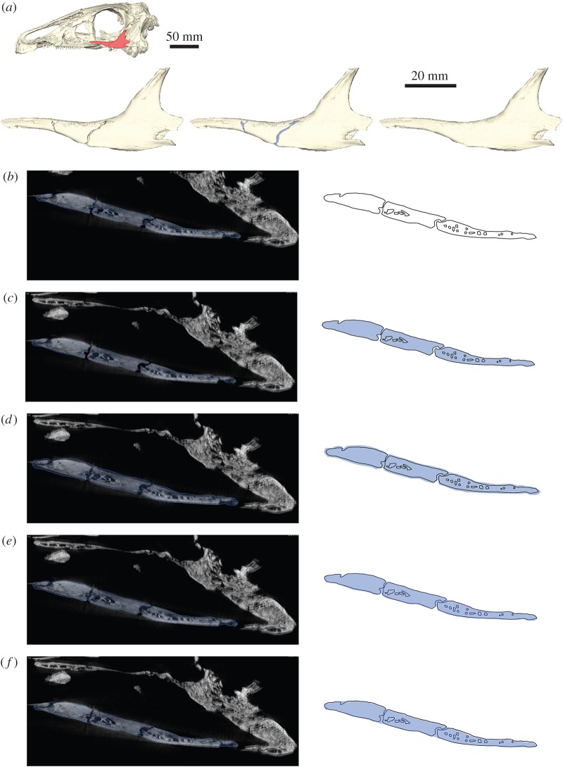
Figure 1.
Removal of breaks and cracks in CT-derived data. (a) Digital representation of the left jugal of Erlikosaurus andrewsi from left to right as originally preserved, with in-filled breaks and fully restored element. Skull image at the top shows position of figured element. (b) CT slice of segmented jugal based on automatic threshold, (c) after hole-filling algorithm, (d) after grow operation, (e) after subsequent shrink operation and (f) manually filled-in breaks. Blue silhouette indicates segmented region according to each operation. All steps performed in AVIZO.

