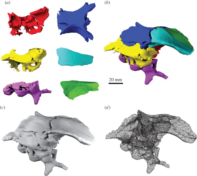
Figure 6.
Manual repositioning of elements. (a) Surface models of disarticulated braincase elements of Dysalotosaurus lettowvorbecki based on CT scanning and photogrammetry. (Red: left laterosphenoid, prootic and opisthotic; yellow: right laterosphenoid, prootic and opisthotic; blue: parietal and supraoccipital; purple: basioccipital and parabasisphenoid; cyan: left frontal; green: right frontal.) (b) Surface model of articulated braincase. (c) Remeshed surface model and (d) polygon mesh. Manual repositioning performed in BLENDER.

