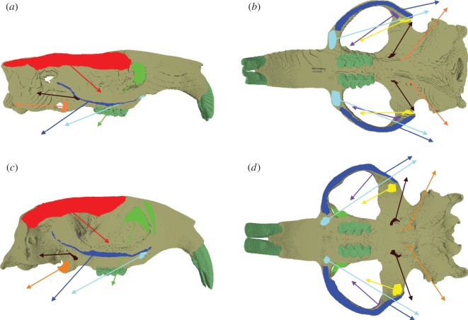
Figure 1.
Attachment sites and vectors of pull of the masticatory muscles in models of Bathyergus suillus, in (a) right lateral and (b) ventral view and Fukomys mechowii, in (c) right lateral and (d) ventral view. Colours of muscle origins and vectors: temporalis, red; superficial masseter, cyan; deep masseter, royal blue; infraorbital ZM, green; anterior ZM, purple; posterior ZM, yellow; lateral pterygoid, brown; medial pterygoid, orange.

