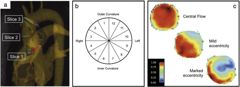
Fig. 1.

Methodological schematic: a) position of the analysis planes in the ascending aorta on the level of the sinotubular junction (S1), mid-ascending (S2) and distal ascending aorta (S3). b) Cross-section of the ascending aorta that describes the distribution of the segments along the aortic wall circumference. c) Peak systolic flow map at the level of the mid-ascending aorta demonstrating central systolic flow, mild and markedly eccentric systolic flow.
