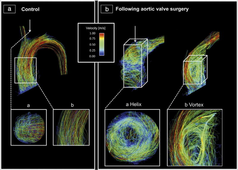
Fig. 3.

Visualization of the blood flow in the ascending aortic using streamlines during peak systole: A: Healthy volunteer with cohesive systolic streamlines with mild helical (a) and no vortical (b) flow. B: Two exemplary cases with AVR, who exhibited helical (a) and vortical (b) flow each graded as severe. a) A mechanical prosthesis (St. Jude Medical 21); b) a stented bioprostheses (Medtronic Freestyle 25). The helical flow is shown in a transverse cut plane, whereas the vortex is shown in a sagittal cut plane.
