Fig. 1.

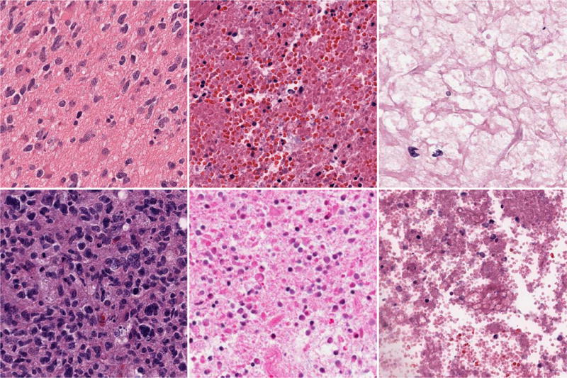
GBM Examples. First column: Tumor; Second column: Transition to necrosis; Third column: Necrosis. Note that the phenotypic heterogeneity is highly diverse in each column.

GBM Examples. First column: Tumor; Second column: Transition to necrosis; Third column: Necrosis. Note that the phenotypic heterogeneity is highly diverse in each column.