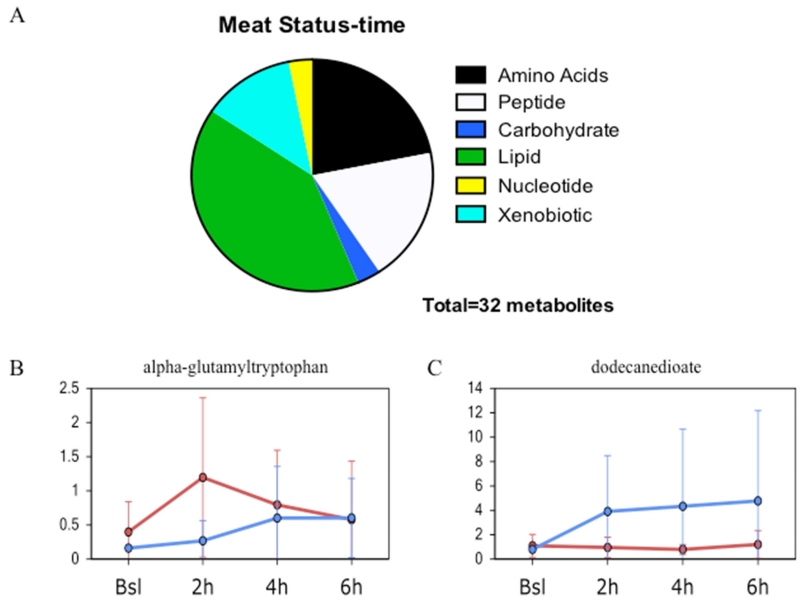
Figure 2. Time-dependent differences between meat allergic and control subjects following meat challenge.
A. Metabolites differentially expressed at baseline showing interaction between meat allergic status and time after challenge with major metabolic pathways and proportion altered displayed. B. Representative line plot showing the time dependent change in expression of the peptide alpha-glutamyltrytophan that showed significant interaction (p<0.05). C. Representative line plot showing the time dependent change in expression of the lipid dodecanedioate that showed significant interaction (p<0.05). The blue line is control and red line meat allergic with the circle representing the mean value and the whiskers representing ± one standard deviation.

