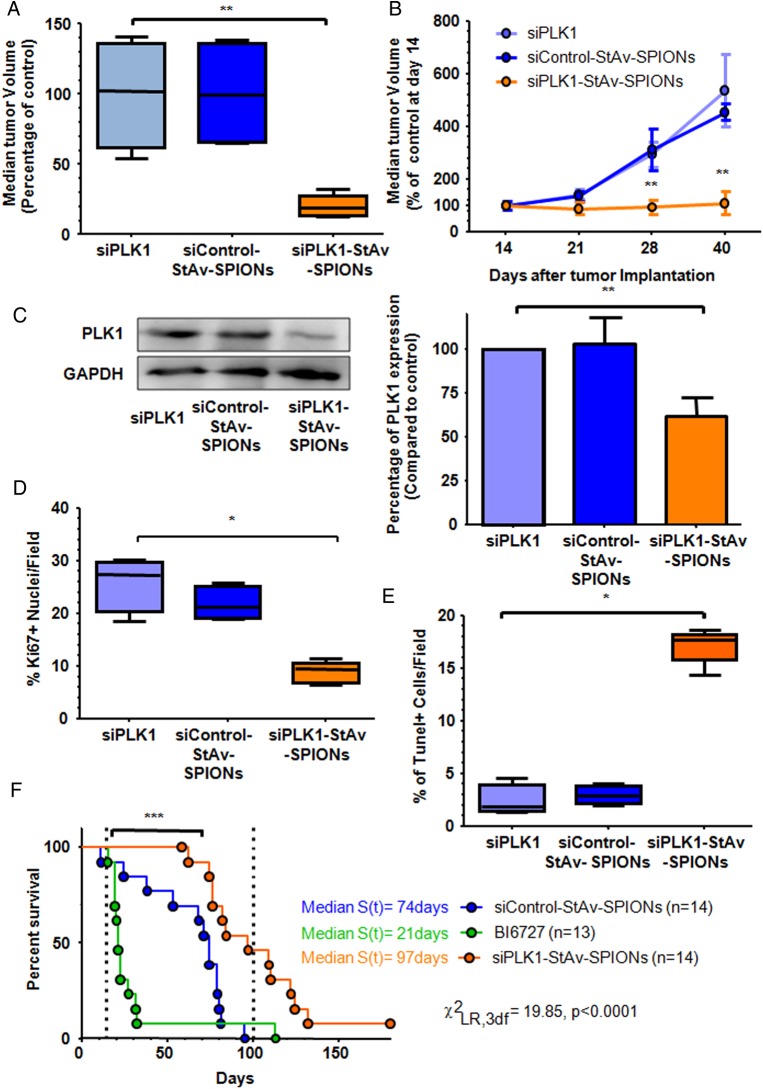
Figure 4.
siPLK1-StAv-superparamagnetic iron oxide nanoparticle (SPIONs) arrest tumour progression in a syngeneic orthotopic tumour model. Tumour-bearing mice were injected intravenously with either siPLK1-StAv-SPIONs, naked siPLK1 or mismatch control siRNA-StAv-SPIONs 14 days after tumour implantation for 3qD over a time period of 4 weeks. (A) Median tumour volume after completion of therapy shows significant reduction in tumour volume of siPLK1-StAv-SPION-treated animals compared with either naked siPLK1 or mismatch-control-siRNA-StAv-SPION (n=5, Kruskal–Wallis test followed by Dunn's multiple comparison post hoc test. H=0.67, p=0.003). (B) In situ tumour measurement using MRI showed stagnancy of tumour growth in the siPLK1-StAv-SPION-treated group over the period of treatment. siControl-StAv-SPIONs or naked PLK1 siRNA did not show any effect on tumour volume (n=3–4, Mann–Whitney U test. U=0.00, p=0.07 (21 days), U=0.00, p=0.002 (28 days), U=0.00, p=0.002 (40 days)). (C) Immunoblotting of PLK1 revealed a decrease in PLK1 expression in the siPLK1-StAv-SPIONs-treated group. Quantification of the expression level showed a significant decrease in PLK1 expression. Data represent mean±SD of three individual experiments. Kruskal–Wallis test followed by Dunn's multiple comparison post hoc test. H=9.91, p=0.007. (D) Quantitative analysis of the percentage of Ki67-positive nuclei in tumour resection specimen proved a significant decrease in proliferation in the siPLK1-StAv-SPION-treated group (n=4–5, Kruskal–Wallis test followed by Dunn's multiple comparison post hoc test. H=9.10, p=0.01). (E) Tunel assay showed a significant increase in apoptotic nuclei upon siPLK1-StAv-SPION treatment (n=4–5, Kruskal–Wallis test followed by Dunn's multiple comparison post hoc test. H=9.09, p=0.01). (F) Kaplan–Meier survival analysis from the time of enrolment to treatment with siControl-StAv-SPIONs (n=14), BI6727 (n=13) or siPLK1-StAv-SPIONs (n=14). Dotted line window indicates maximum duration of therapy. Median survival time of siPLK1-StAv-SPION treatment (96 days) was significantly different to the siControl-StAv-SPION treatment (74 days). ***p<0.001, **p<0.01, *p<0.05.

