
Fig. 7.
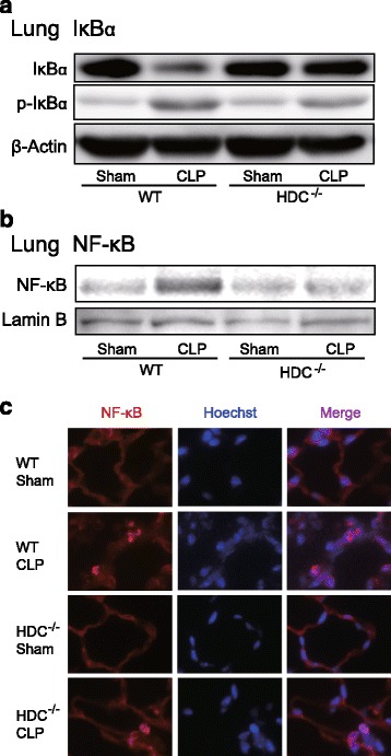
Kinetics of NF-κB activation in lungs of HDC−/− mice following CLP-induced sepsis. Lung tissues were harvested from sham-operated and CLP-induced septic mice 18 h after surgery. a Western blot analysis using anti-IκBα antibody and anti-phospho-IκBα antibody. β-Actin served as loading control. b Nuclear proteins were extracted, and then NF-κB p65 was detected by Western blot analysis. Lamin B served as a nuclear marker. c Immunofluorescent images for NF-κB p65 (red) in lung sections. Nuclei were counterstained with Hoechst 33342 dye (blue). Original magnification, x400. Shown are representative blots from two independent experiments in which the same results were obtained