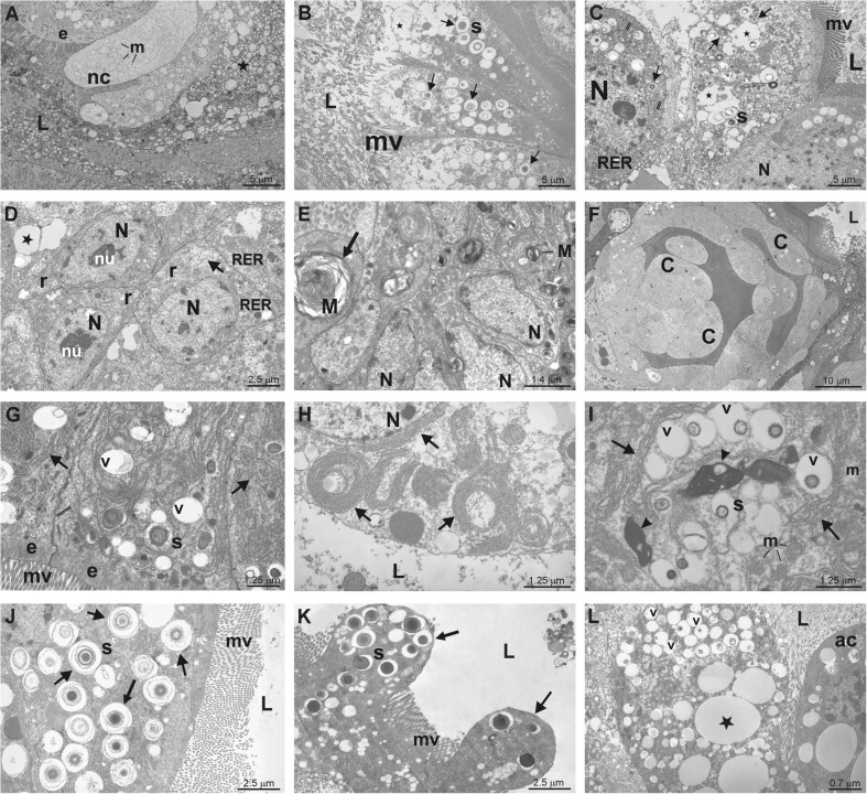
Fig. 3.
Examples of degenerative changes at transmission electron microscope (TEM) level in the midgut cells of Pterostichus oblongopunctatus treated with Cd, Ni or Zn. L – the midgut lumen, mv – microvilli, N – nucleus, nu – nucleolus, stars and v – vacuoles, m – mitochondria, e – epithelial cells, r – regenerative cells in crypts, RER – cisterns of rough endoplasmic reticulum, s – spherites, M – myelinoid body. A: control, epithelial cell (e) neighbours necrotic cells (nc) with swollen mitochondria (m). B: 4th day of Ni exposure, apical portions of epithelial cells containing vacuoles and Ni spherites (arrows) are discharged to the lumen. C: 2nd day of Zn exposure, epithelial cells contain numerous vacuoles and Zn spherites (s). D: 2nd day of Zn exposure, regenerative cells (r) in a crypt showing vacuoles and translucent cytoplasm (arrow). E: 16th day of Ni exposure, an arrow indicates autophagosome (M- myelinoid body, arrow). F: 20th day of Zn exposure, crypt cells (c) are swollen and epithelial cells above crypt cells are degenerated. G: 12th day of Cd exposure, cells contain numerous vacuoles (v) and spherites (s) surrounded by endoplasmatic reticulum membranes (arrows). H: 8th day of Ni exposure, endoplasmatic reticulum forms membranous structures (arrows). I: 12th day of Zn exposure, endoplasmatic reticulum membranes (arrows) surround organelles in autophagosomes, arrowheads indicates residual bodies. J: 4th day of Ni exposure, arrows indicate Ni spherites. K: 8th day of Zn exposure, the budding of epithelial cell cytoplasm with spherites (arrows). L: 8th day of Ni exposure, an epithelial cells with numerous vacuoles and apoptotic cell (ac) discharged to the midgut lumen. Magnification: A, B, C ×1800; D, L ×2800; E, G ×5

