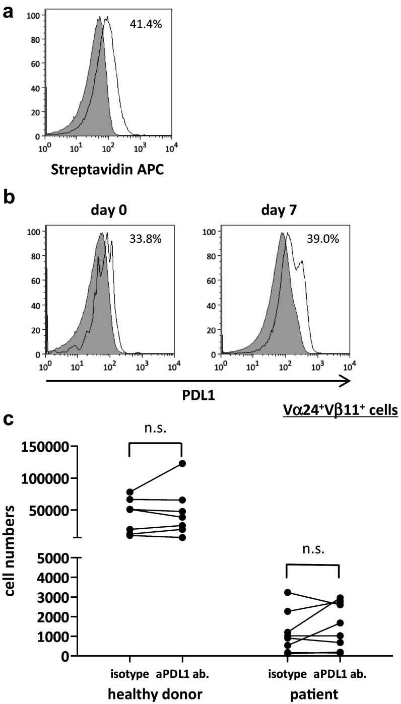
Fig. 2.
Proliferation of human iNKT cells with PDL1 blockade. PBMCs were obtained from six healthy donors and eight non-small cell lung cancer patients. On day 0, PBMCs were stimulated with αGalCer-pulsed IL-2/GM-CSF cultured APCs with anti-PDL1 antibody or isotype control. On day 7, cells were collected and restimulated with PDL1-blocked or isotype control-treated APCs at a ratio of 1:2.5. The cells were collected and counted on day 14, and the proportion of Vα24+Vβ11+ iNKT cells was analyzed using flow cytometry. a Anti-PDL1 antibody binding and PDL1 positivity on APCs were assessed using anti-mouse biotin plus streptavidin staining. b The percentage of PDL1-positive iNKT cells on days 0 and 7 were analyzed with APC-conjugated anti-human PDL1. The gray-shaded histogram represents the isotype control; the unshaded histogram represents PDL1. c The number of Vα24+Vβ11+ iNKT cells on day 7 is shown. PDL1 positivity on APCs was analyzed according to the population comparison method using the FlowJo software program. P values were calculated using the unpaired t test. isotype, isotype control; aPDL1 ab, anti-PDL1 antibody

