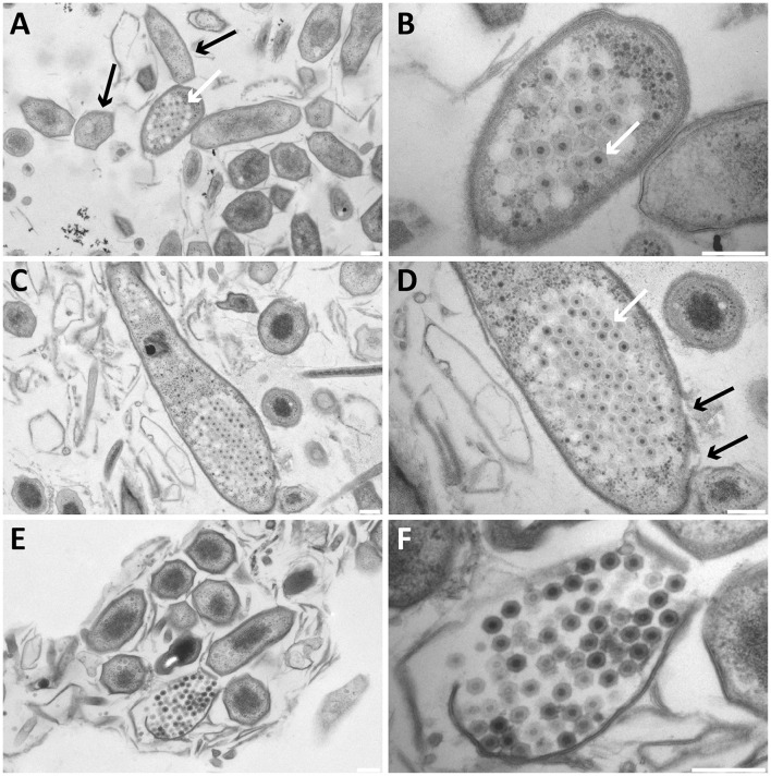
Figure 2.
Transmission electron micrographs of high-pressure frozen, freeze-substituted, resin-embedded, and thin-sectioned Methylomirabilis cells taken from a bioreactor enrichment culture. (A,B) Infected Methylomirabilis cells (white arrow in A) are in clusters among non-infected cells (black arrows in A). The bacteriophage (white arrow in B) has a hexagonal shape and an internal electron dense core. (C,D) The bacteriophages are organized in a highly packed formation (white arrow in D). The replication and assembly of bacteriophages causes the Methylomirabilis cell to swell and eventually the cell wall breaks (black arrows in D). (E,F) Lysed Methylomirabilis cell releasing the viral progeny. (B,D,F) are enlargements of (A,C,E), respectively. Scale bars; 200 nm.

