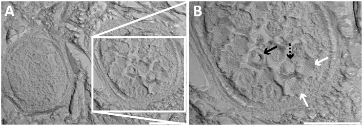
Figure 3.
Transmission electron micrographs of high-pressure frozen and freeze-etched Methylomirabilis cells taken from a bioreactor enrichment culture. (A) Cross-section of a non-infected (left) and infected (right) Methylomirabilis cell. (B) The bacteriophages (white arrows) contain a proteinaceous capsid. The capsid is made of triangular faces built by capsomeres. Concave (dashed black arrow) and convex (black arrow) printing of the internal core is visible in two of the viral particles. Scale bars; 200 nm.

