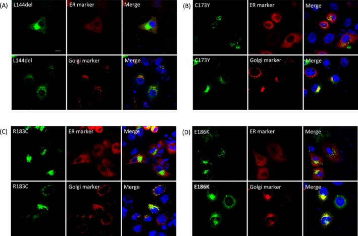
Figure 3.

Subcellular localization of intracellularly trapped GJB1 mutants. Confocal fluorescence images of HeLa cells cotransfected with plasmids encoding GJB1 (labeled with a GJB1‐specific antibody; green) and markers of the ER (DsRed‐ER) or the Golgi (DsRed‐Monomer‐Golgi). Similar staining patterns were observed in cells expressing L144del (A), C173Y (B), R183C (C), and E186K (D); GJB1 staining was mainly observed in the perinuclear region and exhibited colocalization with Golgi markers. Cell nuclei were stained with DAPI (blue). Scale bar, 10 μm.
