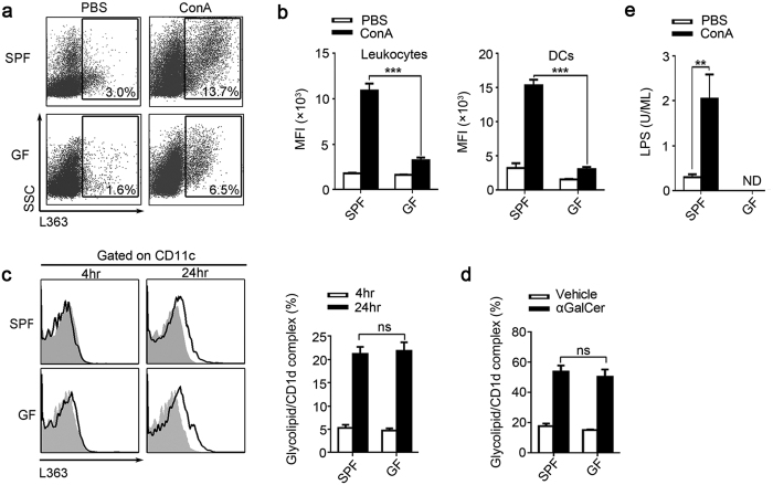
Figure 5. Levels of CD1d-presented and un-presented glycolipids are substantially lower in GF mice.
(a,b) SPF and GF BALB/c mice were sacrificed 24 hr after ConA or PBS injection. (a) Using mAb L363, we detected the percentage of glycolipid/CD1d complexes in the intrahepatic leukocytes, gated on total leukocytes. (b) The mean fluorescence intensity (MFI) of glycolipid/CD1d complexes on total intrahepatic leukocytes and DCs. (c) To compared the glycolipids presenting ability between SPF and GF mice, splenic cells from untreated SPF and GF BALB/c mice were cultured with either αGalCer or vehicle stimulation for 4 and 24 hr. Then used L363 to analysis the glycolipid/CD1d complex expression on DCs by flow cytometry, filled flow cytometric histograms represent cultured with vehicle, and opened histograms represent cultured with αGalCer. (d) Mice were sacrificed 24 hr post αGalCer (250 μg/kg) or vehicle injection. Using L363, the glycolipid/CD1d complex on hepatic DCs was analyzed by flow cytometry. (e) Plasma LPS levels of SPF and GF mice 24 hr after ConA or PBS injection. The data represent means ± SEM (n = 8, two independent experiments), **P < 0.01, ***P < 0.001, ns represents not significant, One-way ANOVA.

