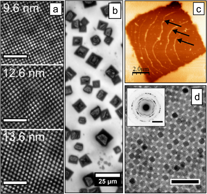
Figure 6.
Self-assembled arrays and mesocrystals formed by the cubic nanoparticles described in this work. (a) High resolution scanning electron microscopy (SEM) images of ordered arrays of nanocubes taken from top-surfaces of self-assembled mesocrystals. Scale bars (white): 100 nm. The images have been FFT-filtered for clarity. (b) Reflected light microscopy images of cuboidal mesocrystals composed of 9.6 nm nanocubes by a conventional drop-casting procedure. (c) Atomic force microscope (AFM) tapping-mode phase image of the surface of a single cuboidal mesocrystal. Growth steps on the crystal surface are highlighted by arrows. (d) TEM image of a multilayer of 9.6 nm nanocubes. Scale bar (black): 50 nm. The inset shows a wide angle electron diffraction pattern of the area. Scale bar (inset): 5 nm−1.

