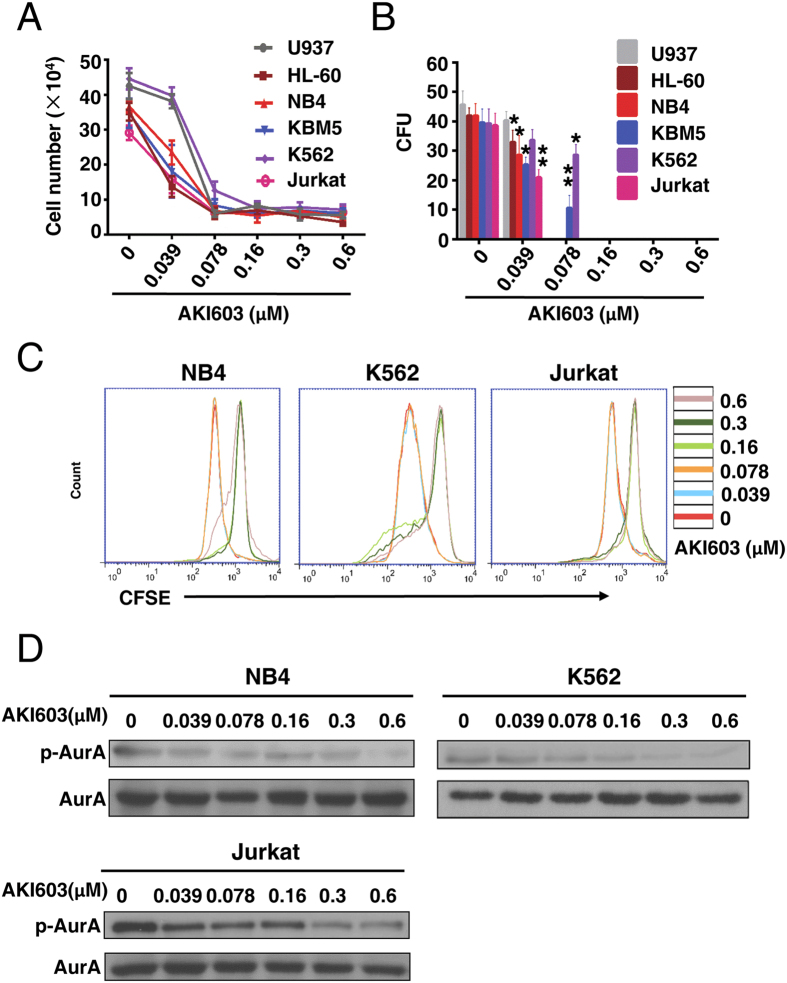
Figure 1. AKI603 extensively inhibits proliferation of leukemia cells.
(A) Leukemia cells were treated with various concentrations of AKI603 (0 μM, 0.039 (0.0390125) μM, 0.078 (0.078125) μM, 0.16 (0.15625) μM, 0.3 (0.3125) μM, 0.6 (0.625) μM) for 48 h. Cell counting assay was performed. The mean values from three independent experiments are presented. (B) The colony formation of cells treated with AKI603 for 10 days were analyzed. The statistical analysis of the colony formation assay is shown (mean ± SD, *p < 0.05, **p < 0.01 vs. 0). (C) NB4, K562 and Jurkat cells were stained by CFSE and treated with various concentrations of AKI603 for 48 h. The levels of CFSE fluorescence were analyzed by flow cytometry. (D) NB4, K562 and Jurkat cells were treated with various concentrations of AKI603 for 48 h. The lysates were subjected for western blot analysis of p-AurA (Thr288) and AurA expression. The data are representative of three independent experiments.

