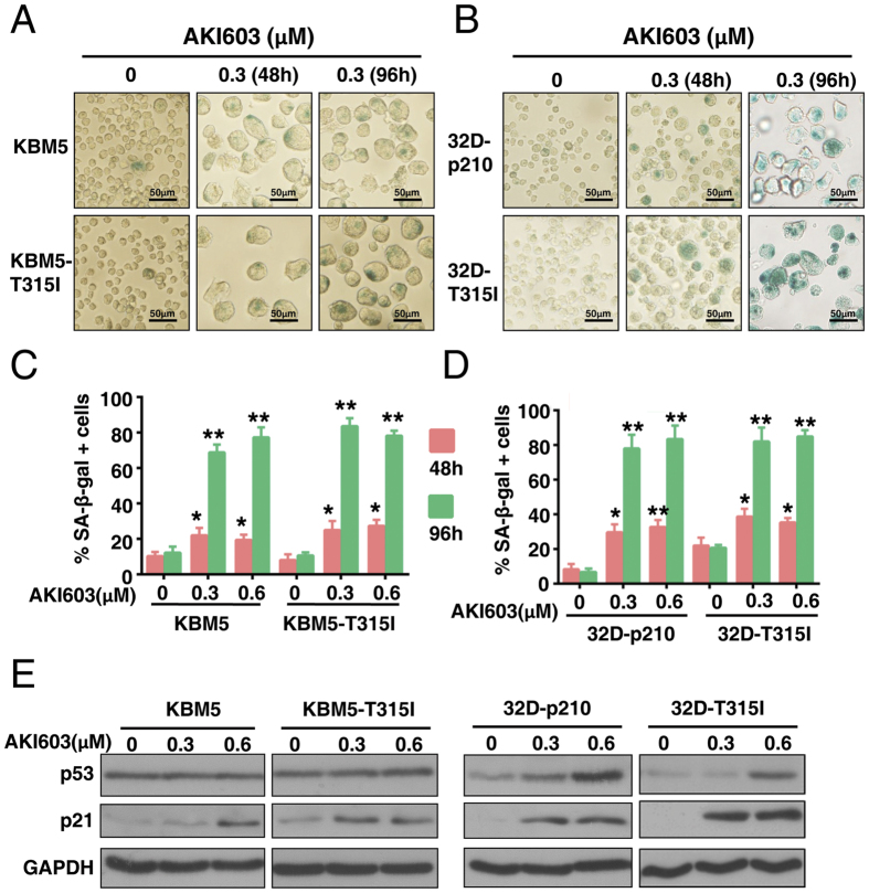
Figure 3. Inhibition of AurA kinase by AKI603 induces cellular senescence.
(A,B) KBM5, KBM5-T315I, 32D-p210 and 32D-T315I cells were treated with 0.3 μM of AKI603 for 48 h or 96 h, and senescence was determined using SA-β-gal staining. (C,D) The statistical analysis of the SA-β-gal assay is shown. More than 300 cells per sample were counted to determine the percentage of senescent cells (mean ± SD, *p < 0.05, **p < 0.01 vs. 0). (E) KBM5, KBM5-T315I, 32D-p210 and 32D-T315I cells were treated with 0.3 μM and 0.6 μM of AKI603 for 96 h. The lysates were subjected to western blot to analyze the expression of p53 and p21. The data are representative of three independent experiments.

