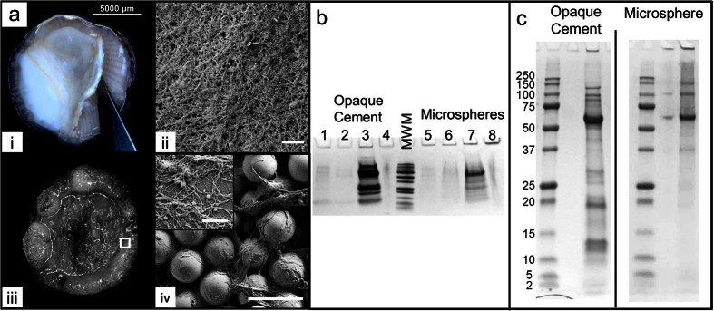
Figure 1. Collection and breakdown of cement samples from adult A. amphitrite barnacles.
(a) Examples of fine nanofibrils networked into dense materials observed in multiple collection methods: (i) Thick and opaque cement layer secreted from the barnacle underside, (ii) SEM micrographs of (i) showing a mat consisting of fine nanomaterials, scale bar represents 1 μm. (iii) Underside of a barnacle settled on a bed of glass microspheres after one day, with large accumulations at the periphery, (iv) SEM micrographs of barnacle-adhered microspheres entrapped by cement secretions, scale bar is 100 μm. Inset, 2.25 μm2 image showing close up of nanofibrils on beads, scale bar is 500 nm. (b) Breakdown of collected cement using ethyl acetate (lanes 1 and 4), urea (lanes 2 and 6), hexofluoroisopropanol (lanes 3 and 7), and dithiothreitol (lanes 4 and 8) solvents, eluted proteins analyzed by SDS-PAGE and (c) Full length PAGE run of ‘opaque’ and ‘microsphere’ adhesives after dissolution by HFIP with an abundant protein released at 63 kDa among other well-known bands at 250, 100, 35, and 19 kDa. Left is ‘opaque’ cement, right is ‘microsphere’. Uncropped whole gels from (b,c) are available in Supplementary Figure S7.

